Neuroimmunology: The Great Brainstorming!
At the Center of Immunology Marseille-Luminy, Réjane Rua and her co-workers are furthering knowledge of the mechanisms that protect our brain. They are studying the properties and functions of newly discovered sentinel cells in our meninges.
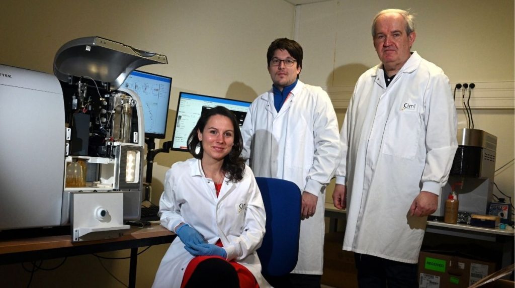
Just 5 years ago, a discovery was made that has profoundly modified our understanding of brain protection: the meninges, the membranes that surround and protect the cerebral cortex, are not just a purely structural mechanical support… they are also host to myriad sentinel cells of the immune system! The brain and immune system, historically considered to be distinct, are actually closely linked. The meninges have been found to constitute an essential interface of the central nervous system, serve as supports for brain development, be necessary for brain balance, and play a decisive role in a variety of diseases. A better understanding of meningeal immunity could then reveal therapeutic targets in neurological conditions, such as Parkinson’s disease. However, the researchers have only just begun to explore this very young field that is neuroimmunology. The mysteries of the various immune cell populations found in the meninges remain to be elucidated, and their roles understood. Thanks to an ERC Starting Grant, the Center of Immunology Marseille-Luminy has recently acquired Europe’s first Cytek spectral cell sorter. This ultra-precise machine capable of analyzing around fifty cell markers simultaneously will enable Réjane Rua* and her team to illuminate these fascinating meningeal guardians as never before.
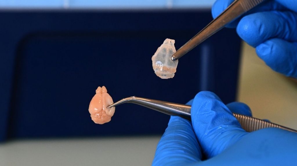
The meninges, located between the brain and the braincase, contain more varieties of immune cells than the brain as a whole! They constitute a major immunological barrier in which macrophages, a subtype of white blood cells, constantly monitor the vessels to know what is going in and out… It is this type of cell that interests Rua: “We want to understand why they are there, what role they play in the brain’s growth and protection against pathogens. To do so, we need to explore their diversity.” Each type of immune cell has its own specific surface markers. Rua uses a panel of different fluorescent dyes (fluorophores) to recognize these markers and distinguish between the cells. Composed of an antibody directed towards a molecule of interest that is attached to a fluorescent particle, each fluorophore emits a distinct light following excitation by a specific wavelength.
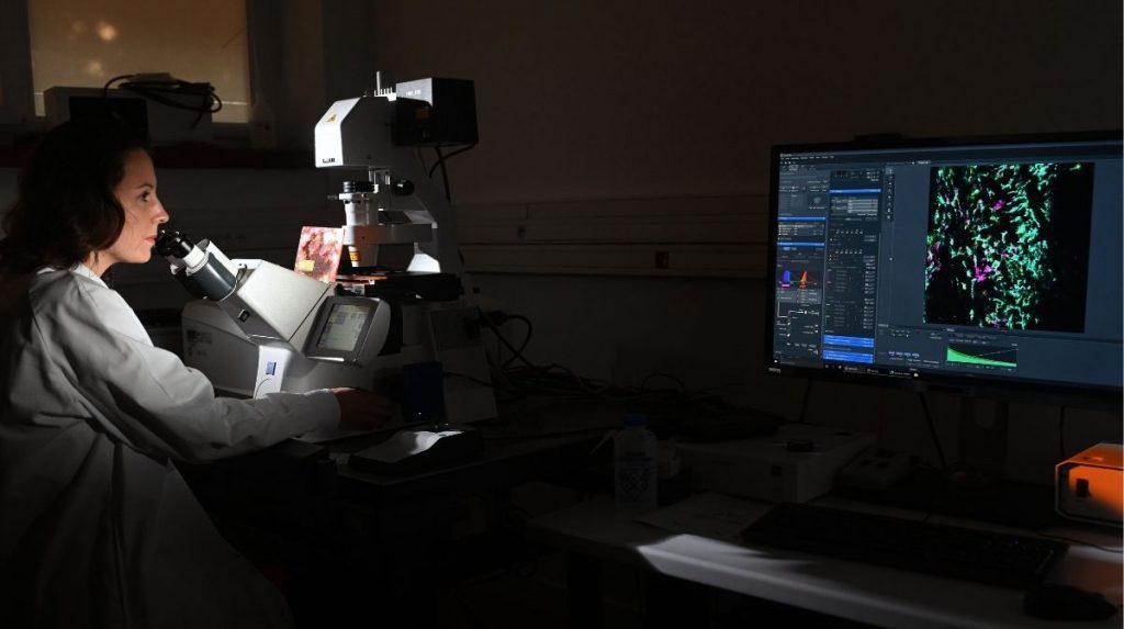
This fluorescence is visible under a confocal microscope that can distinguish up to 10 light spectra. The location of the cells in the meninges provides indications regarding their functions. In particular, there is an abundance of macrophages (inactivated in blue and green, activated in pink) around the vessels supplying the brain. Having sentinels at these locations is strategic in order to monitor, identify and block pathogens before they invade the brain.
Spectral cytometry is a fluorescence analysis technique that complements microscopy. It measures, distinguishes, counts, analyzes, and characterizes each cell at high speed, according to more numerous criteria. Rua’s team has been equipped with Europe’s first Aurora CS spectral cell sorter, which combines the capacities of conventional cytometry analysis with unparalleled accuracy, distinguishing up to 50 different spectra. “This machine enables an incredibly more accurate and in-depth characterization of the cell populations,” explains the researcher.
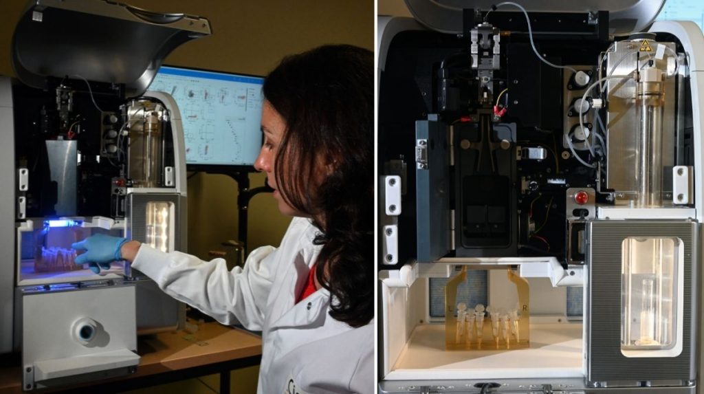
Each fluorophore is attached to a different marker, the combination of which characterizes a cell subtype. Rua can therefore differentiate the macrophages, which are large cells capable of digesting foreign bodies that enter our body, from other immune cells, but also the subtypes of macrophages and their particularities. “This is the first time we have a resolution that makes it possible to sort these various cell populations. We really have the possibility to know who is doing what in the meninges!” smiles the scientist.
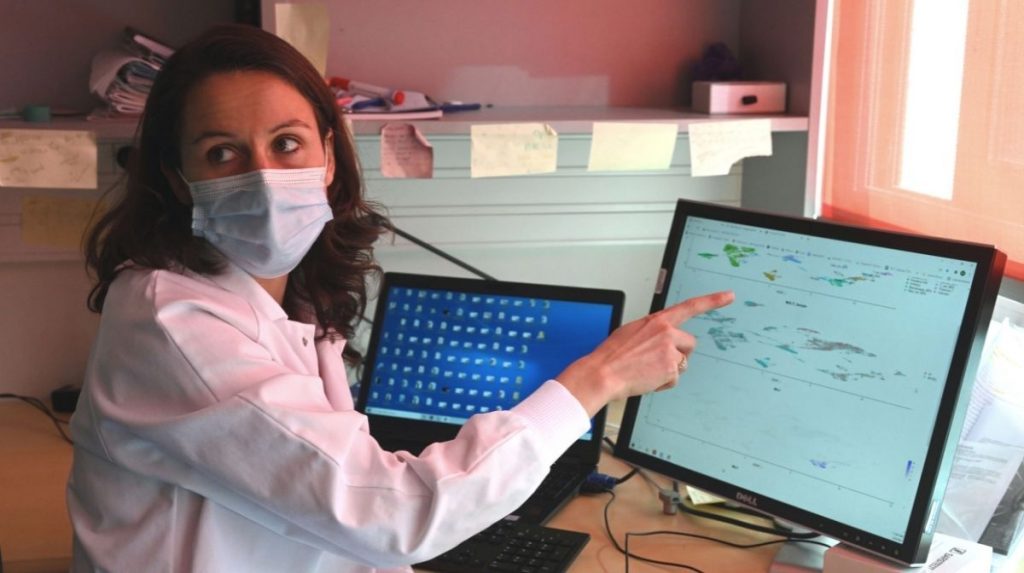
On the sorted cells, Rua looks at the various RNA produced by a cell in order to identify the genes activated in the subpopulations. From these it is possible to deduce properties and therefore functions. If antiviral RNA is found, it can be assumed that the macrophage defends itself against viruses, for example. The presence of growth factor RNA suggests a role in the growth and survival of neurons, and therefore in the development of the brain.
These discoveries feed into different projects being worked on by the team, which seeks to understand how the meninges detect and block viruses that try to enter the brain (neuroinfection), how these meninges promote brain growth (neurocognition), and how they support skull growth (neurobiomechanics). “We are at the very beginning of the journey, specifies Rua. We still need to understand how it all works! Then we will be able to consider interventional therapies.”
Note:
* unit 1104 Inserm/CNRS/Aix-Marseille Université, Immunosurveillance of the central nervous system team
